
single tooth implant before and after
Single Tooth Implant Before and After: How to Read Results Realistically
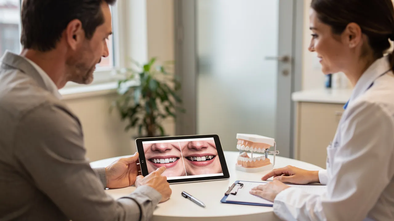
What a single-tooth implant before-and-after can—and cannot—tell you
A single-tooth implant before-and-after comparison can help you judge visible changes, but it cannot establish that an implant is appropriate for your mouth or predict that you will receive the same result. Use the images as a starting point for better questions.
A useful review follows three steps:
- Identify what treatment the images show and which stage the after image represents.
- Compare the photographs under similar conditions, then inspect the tooth, gum line, surrounding teeth, and visible smile as a group.
- Take unanswered questions to an examination and treatment-planning discussion rather than choosing treatment from photographs alone.
A single-tooth implant may involve an implant placed in the jawbone, a healing period that includes osseointegration, and a custom crown attached after integration. The crown is the part you usually assess in a smile photograph; the implant itself is part of the supporting treatment below the visible tooth. A photo can therefore show an appearance at a particular stage without showing the examinations, imaging, healing, or planning behind it.
The practical boundary is specific: use photographs to discuss appearance, not to determine what is happening beneath the gum line, whether the implant has integrated, or which replacement path fits your situation. Those questions require an individualized dental assessment.
Decode the replacement shown in the images
A single-tooth implant replacement has more than one part, so start by identifying what you can actually see. The visible after image generally represents the custom crown that replaces the missing tooth’s visible portion. The implant is placed in the jawbone as a foundation, and healing allows osseointegration—the implant’s fusion with the jawbone—before the crown is attached.
That distinction changes how you read the comparison. If the before photograph shows a space, the after photograph may show a tooth-shaped crown that fills the visible gap. The photograph does not show whether the implant has integrated, how the site was evaluated, or what conditions shaped the treatment plan. Those questions belong in the clinical discussion.
Look for a caption or case description that identifies the treatment type. A gallery may place different procedures next to each other, including:
- A single implant-supported crown.
- A bridge supported by adjacent teeth.
- A full-arch restoration.
- A temporary or same-day provisional rather than a final crown.
The label matters because the apparent change in the photograph may come from a different treatment design than the one you are considering. A bridge and an implant-supported crown are not interchangeable examples, even when both replace a single visible tooth. Ask what was placed, which teeth were involved, and whether the image shows a temporary stage or the final restoration before drawing comparisons.
Also separate the visible crown from the parts you cannot see. A case caption may identify the final restoration, but the image alone cannot tell you how the implant was positioned, what imaging supported the plan, or how healing progressed. Record those as questions rather than filling in the gaps from the photograph.
Verify that the comparison is genuinely comparable
A comparable before-and-after pair gives you a more useful view of visible change than two photographs taken under different conditions. Similar camera positions and similar lighting make differences easier to judge; a change in angle, framing, or lighting can make a restoration appear more or less noticeable than it is.
Before studying the tooth, check the image conditions:
- Are the before and after views taken from a similar angle and distance?
- Is the mouth open to a similar degree, with the lips in a similar position?
- Does the lighting show the teeth and gum line clearly in both images?
- Are both images from the same stage of treatment, or is one an early provisional?
- Does the caption identify the tooth position and the type of final restoration?
A caption may also identify healing, grafting, treatment stages, or the restoration type. That information gives the photograph context that the image itself cannot provide. If the caption is brief, write down the missing details instead of assuming that an attractive after image represents the finished result.
A consistent pair still has limits. You can compare visible position, proportion, shade, and gum contour, but you cannot infer whether the implant has integrated or whether the treatment was suitable for the person shown. You also cannot assume that a different tooth position, gum shape, neighboring-tooth condition, or healing course will produce the same appearance for you. Treat image consistency as a condition for fair visual comparison, not as proof that the clinical circumstances are alike.
If the before and after views use different smiles, ask how much of the apparent change may come from the photograph itself. That question does not dismiss the example. It keeps your conclusion limited to what the paired images can actually show.
Find out whether the result is temporary or final
A provisional restoration can look favorable while the gums and implant are still healing, so an attractive early photograph is not automatically the final result. Identify the treatment stage before you use the image to set expectations.
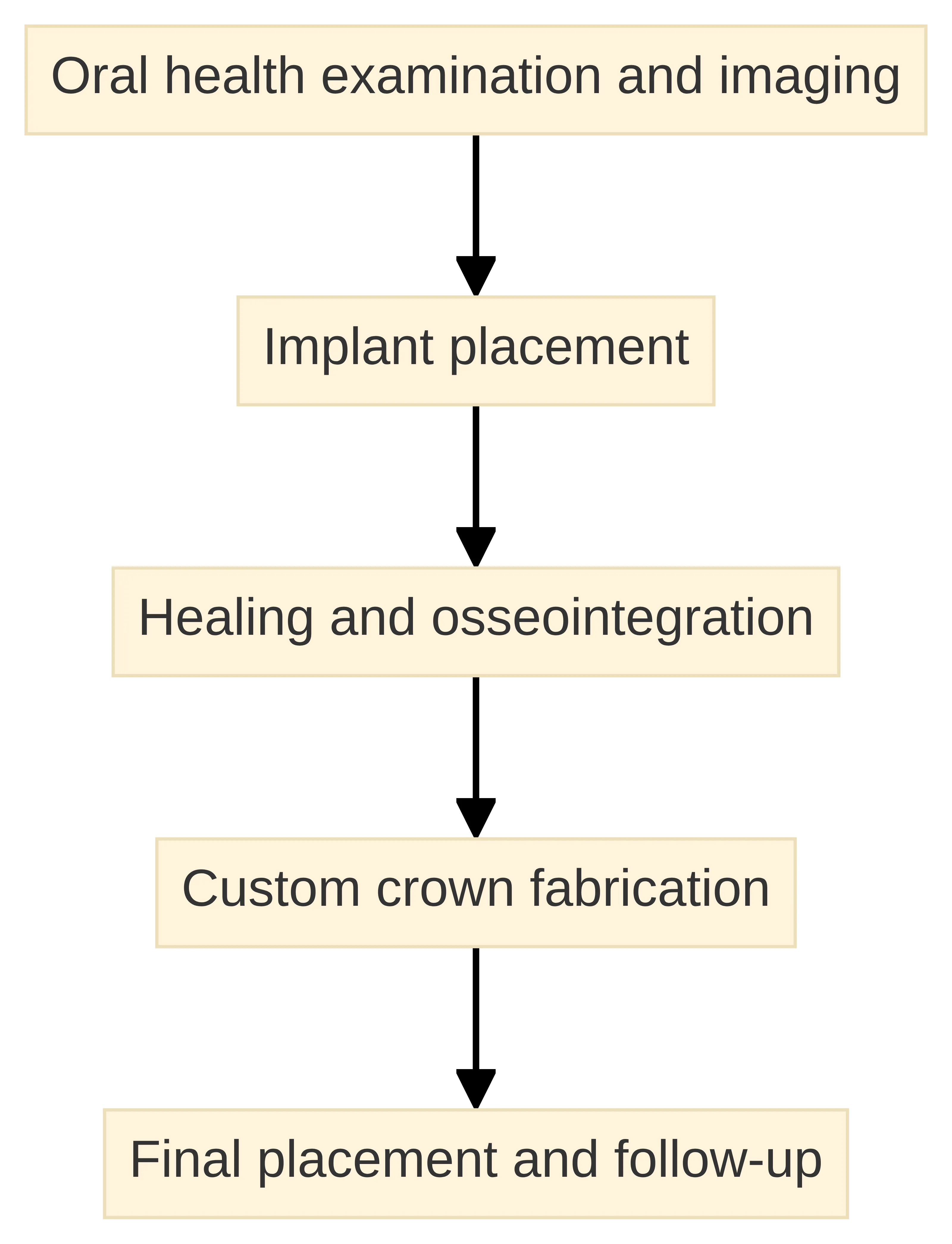
A sequence described for a single-tooth implant can include an oral-health examination and imaging, implant placement, healing and osseointegration, custom crown fabrication, final placement, and follow-up. The sequence provides a map for your questions, not a personal timetable. Your evaluation may require discussion of the site, the proposed restoration, and the conditions that affect staging.
Use the case description to separate these stages:
- Initial or pre-treatment image: This may show the missing tooth or the condition before treatment.
- Provisional image: This may show a temporary tooth while the gums and implant continue healing.
- Final-restoration image: This should be identified as the completed crown rather than assumed to be final because it looks polished.
- Follow-up image: This may show the restoration after additional healing or review, if the case description provides that information.
Ask whether the after image was taken immediately after placement, during provisional use, or after the final crown and follow-up. Ask what healing stage the image represents and whether the visible gum contour was still changing at that point. The point is not to reject an early image; it is to understand exactly what it can show.
The same question applies to your own proposed plan. Ask which stages you would complete, which component would be temporary, what follow-up would assess, and what would need to happen before a final crown was placed. A polished provisional may help you visualize direction, but it should not be treated as a promise about the completed result.
Inspect the visible result systematically
A visible implant result is easier to assess when you inspect the same features in the same order rather than reacting to the overall smile. Use the checklist below to turn an impression into specific questions.
- Tooth position: Does the replacement occupy the expected space, and does it appear aligned with the neighboring teeth? A front-to-back or side-to-side shift may change the way the smile looks even when the crown fills the gap.
- Gum contour and symmetry: Does the gum line around the replacement appear consistent with the adjacent teeth? Look for the height and shape of the tissue rather than treating a smooth-looking photograph as proof of the underlying condition.
- Color and surface appearance: Does the shade appear compatible with the surrounding teeth under the same lighting? A photograph can alter shade, so ask how color was selected and whether the displayed image shows the final crown.
- Shape and proportion: Does the crown’s width, length, contour, and visible surface fit the neighboring teeth? A restoration can fill the space while still requiring discussion of proportions and design.
- Bite and function: A frontal image cannot show how the teeth meet when you close or move your jaw. Ask how the proposed crown would be assessed for function rather than treating a good smile photograph as evidence about your bite.
- Surrounding teeth: Does the replacement look compatible with the teeth on both sides? Inspect their position, color, shape, and gum line too, because the surrounding teeth frame the result.
- Whole-smile context: Compare gum symmetry, tooth proportions, lip support, and the way the restoration fits the surrounding teeth. These features can matter more than the isolated crown.
Write down what you can see and what you cannot. For example, you may be able to say that the crown appears proportionate in a consistent image, while you cannot determine whether the implant has integrated or whether the bite is comfortable. That separation keeps an appearance judgment from becoming a treatment conclusion.
Use the same order when you review your own proposed design. First ask whether the replacement fills the space naturally. Then ask how its gum contour, color, shape, bite, and relationship to adjacent teeth will be assessed. A checklist gives you a clearer conversation without turning a photograph into a diagnosis.
Look beyond integration to the esthetic and functional context
Implant integration and a pleasing visible result are related but separate questions. The cited literature explains that successful osseointegration does not always correspond to a successful esthetic outcome. Implant position and the surrounding soft tissue can influence whether the restoration appears esthetic and functional.
That distinction explains why the crown alone is not the whole story. A photograph may show a tooth that appears natural in color and shape, while the treatment discussion still needs to address:
- How the implant position relates to the planned crown.
- How the gum contour around the site will be assessed.
- How the restoration will fit the neighboring teeth and the way your teeth meet.
- Whether the proposed design is temporary or final.
- What the examination and imaging show about the site and surrounding oral health.
The visible result also depends on context outside the crown. The position of the missing tooth, the condition of adjacent teeth, and the shape of the gum line can change what a replacement needs to accomplish visually. Ask how those features affect the design rather than asking for a promise that the crown will look identical to a photograph.
An implant that has integrated is not automatically an esthetic guarantee, and a photograph that looks attractive is not evidence of integration. Keep those two judgments separate. Ask what has been examined, what the imaging contributes, and how the proposed restoration will be assessed for both appearance and function.
This distinction is especially useful when a gallery emphasizes the finished crown. The visible tooth may be the part you care about most, but its appearance is connected to the position of the implant and the surrounding tissue. Ask how the proposed design accounts for that relationship instead of evaluating the crown in isolation.
Treat variation as a reason for evaluation, not a promise or warning
Variation between implant photographs is a reason to ask better site-specific questions, not a reason to promise the displayed result or assume that a different result would be a failure. Treatment options depend on diagnosis, oral-health status, needs, and preferences, while implant planning may involve examination and imaging.
For your evaluation, ask how these factors could shape the plan:
- Implant site: Is the tooth in a visible part of the smile, and what does that position require from the crown and surrounding tissue?
- Bone and gum conditions: What does the examination or imaging show at the site, and would those findings affect the proposed sequence or restoration design?
- Healing: Which parts of the appearance may change while healing and osseointegration continue?
- Restoration design: How will the crown’s shade, shape, contour, and contact with neighboring teeth be planned?
- Adjacent teeth: Are the teeth beside the space suitable to remain as they are, or would an alternative involve preparing them?
- Your priorities: How do appearance, function, treatment stages, recovery questions, and cost affect the options you want to compare?
The evaluation should turn general uncertainty into specific information. You do not need to decide from a gallery whether an implant will suit you. You do need to know what examination, imaging, and diagnosis will be used to discuss candidacy and alternatives.
If a proposed result is described with certainty before those details are assessed, slow the conversation down. Ask what is known from your records, what remains uncertain, and which part of the plan addresses each concern. If you are uncomfortable with a recommendation, asking about alternatives or seeking a second opinion is a reasonable next step.
A useful comparison is therefore not “Which photograph do I like most?” It is “Which questions does this photograph raise about my site, the proposed restoration, and the alternatives?” That shift protects you from treating another person’s visible result as a forecast.
Compare the implant with the other single-tooth replacement path
Replacing one missing tooth may involve an implant-supported crown or a three-unit fixed partial denture, and the better option depends on the case. A before-and-after image cannot decide between them because the choice depends on diagnosis, oral-health status, needs, and preferences.
Use the comparison below to organize the discussion rather than rank either option universally.
| Question | Implant-supported crown | Three-unit fixed partial denture |
|---|---|---|
| What should you identify in the plan? | Ask how the implant, abutment, and crown would replace the missing tooth and which stages are proposed. | Ask how the bridge would replace the missing tooth and which adjacent teeth would support it. |
| What should you ask about the adjacent teeth? | Ask whether the neighboring teeth would remain untreated under the proposed plan. | Ask whether the two adjacent teeth would need preparation for the fixed partial denture. |
| What should you ask about the site? | Ask how the examination and imaging would affect implant position, healing, and crown design. | Ask how the condition and position of the supporting teeth affect the bridge plan. |
| What should you compare financially? | Ask what the estimate includes at each stage and what your insurance plan may contribute. | Ask the same questions so the estimates cover comparable parts of treatment. |
| What should you compare after treatment? | Ask about follow-up, care, function, appearance, and the issues that would require review. | Ask about follow-up care for the bridge, its function and appearance, and issues that would require review. |
The central trade-off is not simply “implant versus bridge” in the abstract. It is how each proposed path addresses your missing-tooth site, neighboring teeth, oral-health findings, priorities, and financial boundaries. The professional discussion should explain why a particular plan fits the findings and what alternatives would change.
Ask for both options when both are clinically relevant. Then ask what would make one option unsuitable, what preparation each would require, and how each would affect the teeth around the space. A comparison is useful only when the plans are specific enough for you to compare their stages, conditions, care, and costs.
A photo gallery may show only one path, but that does not make the pictured path the only path available for your situation. Use the image to discuss the appearance you want to understand, then ask how an implant-supported crown and a fixed partial denture would address the same missing-tooth space.
Turn the images into an evaluation question list
An evaluation question list turns a photograph into a focused conversation. Bring the image or page that prompted your questions, but ask about your own findings rather than asking for the pictured result.
Use these groups of questions:
About candidacy and the site
- What does my oral-health examination show at the missing-tooth site?
- What imaging is needed, and what will it help you assess?
- How do the bone and gum conditions at this site affect the proposed plan?
- What findings would change the sequence or make another replacement worth discussing?
About the proposed restoration
- Is the proposed replacement an implant-supported crown, a bridge, or another restoration?
- Which parts are the implant, abutment, provisional, and final crown?
- How will the crown’s position, shape, color, and gum contour be planned?
- How will you assess the bite and the fit with my neighboring teeth?
- Is the example I saw a temporary result, a final restoration, or a follow-up image?
About stages and healing
- What are the stages from evaluation through final placement?
- Which stage includes implant placement, and which stage includes healing and osseointegration?
- What follow-up visits are part of the proposed plan?
- What changes should I expect while healing, and what should I ask about if the course differs from the plan?
After final placement, ongoing care includes following the provided care instructions and attending follow-up visits. These visits are an opportunity to discuss healing, the restoration, and the surrounding tissue over time.
About ongoing care and risks
- Which care instructions apply during healing and after final placement?
- Which follow-up visits are planned, and what will be checked at each one?
- Which risks or complications are relevant to my site and proposed plan?
- How could those issues affect appearance, function, healing, or the treatment sequence?
- Which changes should prompt me to contact the dental team?
About alternatives
- What treatment options could address this space?
- Would an alternative require preparation of the adjacent teeth?
- How do the alternatives compare for appearance, function, stages, care, and cost in my situation?
- What would you recommend based on the diagnosis, and what would make another option reasonable?
Treatment options should be discussed through your diagnosis, oral-health status, needs, and preferences. A question list helps you hear that explanation clearly without treating a photo as a diagnosis.
Save the questions that the image cannot answer separately from the observations you can make. For example, “the crown appears lighter than the neighboring tooth” is an observation; “will the final shade be selected to match my surrounding teeth?” is an evaluation question. Keeping those categories separate makes the appointment more efficient.
Ask for a complete planning and financial picture
A complete implant discussion includes the clinical plan, the treatment sequence, the financial estimate, insurance questions, and follow-up care. Do not compare a photograph or a headline price without knowing which stages and components are included.
Ask the dental team to explain:
- The examination and imaging planned before treatment.
- The proposed implant position and restoration design.
- The stages from placement through healing, abutment, crown, and follow-up.
- Any provisional restoration and what would lead to final placement.
- The care and follow-up expected after each stage.
- The risks and limitations that apply to the proposed plan.
- The total estimated cost, the cost of each stage, and what is excluded.
- What your dental insurance plan may cover and what patient responsibility may remain.
- Whether the alternative treatment has a different sequence or financial structure.
We also handle implant placement, the abutment, and the final crown in-house. Those are descriptions of our process, not a prediction of your result. Ask how the documented planning process would apply to your proposed case and what it would show about your site.
We provide verification of benefits before treatment. That step can organize the insurance conversation, but you should still ask what the estimate includes, what your plan may contribute, and what remains your responsibility. The dental diagnosis and treatment discussion should come before treating financing as the reason to choose one option over another.
A cost question is clearer when you divide it by stage. Ask separately about evaluation and imaging, implant placement, any provisional restoration, the abutment, the final crown, follow-up, and the alternative treatment. Then ask which items are included in the estimate and which may be separate. This gives you a basis for comparing plans without assuming that two prices cover the same work.
Ask how the proposed plan connects appearance, function, healing, follow-up, and cost. A financial figure without that clinical context cannot tell you whether the plan matches your needs.
Bring the right information and request an evaluation
A dental implant evaluation is the appropriate next step when you want to move from a photograph to information about your own missing-tooth site. Bring your insurance card, a list of current medications, and prior dental records or X-rays to your first visit at Westborough Dental Care. Bring your saved image questions too.
Use the appointment to ask what the examination and imaging show, which replacement options fit the diagnosis, how the stages and healing would be handled, how the crown would be planned for appearance and function, and what the complete financial picture includes. A qualified dental professional must determine individual suitability through examination, diagnosis, and treatment planning; this guide cannot make that decision for you.
A careful before-and-after review has done its job when you can describe what the image shows, identify what remains unknown, and ask a focused question about the next decision. You do not need to promise yourself an implant based on a photograph. You need a plan that connects your findings, alternatives, priorities, care, and costs.
Take the evaluation one question at a time. Ask what the images can show, what your examination adds, which alternatives are relevant, what each stage involves, and how the estimate and insurance discussion apply to the proposed plan. That is a more reliable next step than treating another person’s result as a forecast for you.
What should you confirm before scheduling a single-tooth implant evaluation?
What does a single tooth implant replace?
A single tooth implant replaces the visible portion of a missing tooth with a custom crown, supported by a titanium implant placed in the jawbone.
How can I assess a before-and-after example of a single tooth implant?
To assess a before-and-after example, compare the tooth position, gum contour, color, shape, bite, and the relationship to surrounding teeth, ensuring the images are taken under similar conditions.
What factors can affect the results of a single tooth implant?
Results can vary based on the implant site, bone and gum conditions, restoration design, healing process, and the condition of neighboring teeth.
What questions should I ask during a dental implant evaluation?
During an evaluation, ask about treatment alternatives, imaging or digital planning, treatment stages, estimated costs, insurance coverage, and follow-up care.
How does Westborough Dental Care ensure precision in dental implant placement?
Westborough Dental Care uses digital planning with an in-house Cone Beam CT scanner and places implants using surgical guides for higher precision.
Need help applying this?
Talk to a billing specialist who knows your payer mix.
Westborough Dental Care welcomes new patients in South San Francisco. Book a visit to meet Dr. Zoulas and the team.
Schedule a meeting