
pictures of dental implants that failed
What Pictures of Dental Implants Can and Cannot Show
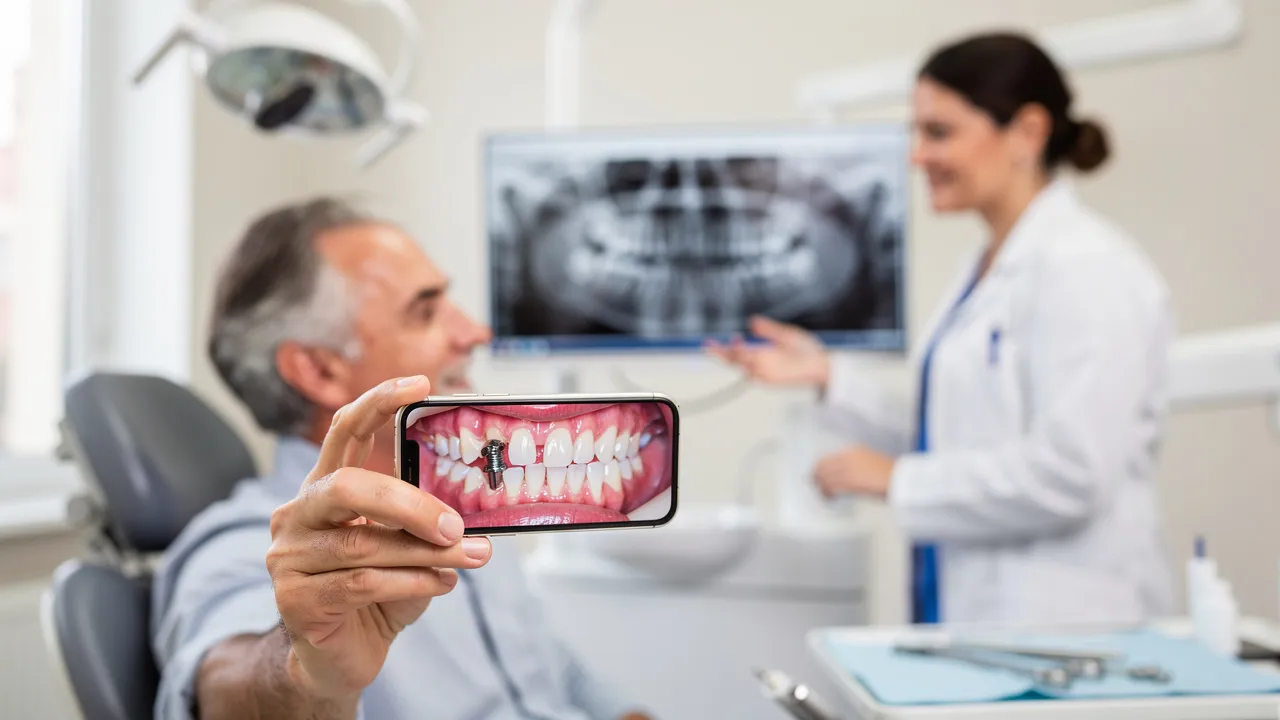
Pictures can raise questions but cannot confirm implant failure
Dental implant photographs can help you identify a change worth discussing, but they cannot confirm that an implant has failed. An image may show the gum around the restoration, a change in color or shape, or a visible part of the crown. It cannot show every finding beneath the gum, establish whether the implant is mobile, or replace an examination and possible imaging.
Implant concern can feel urgent when a tooth looks different, the tissue appears swollen, or the crown seems out of place. A photograph can give you a useful starting point without giving you a diagnosis. Use it to describe what changed, when you noticed it, and whether the concern is accompanied by pain, bleeding, swelling, or movement.
An image also freezes one moment. It cannot tell you whether a change is new, stable, or worsening unless you provide that timeline. Earlier photographs and symptom notes can add context, while the clinical meaning still requires an assessment.
The useful way to read an image is as a prompt for questions:
- What part of the implant restoration is visible?
- What tissue change, if any, can you actually see?
- What symptoms or stability concerns cannot be judged from the image?
- What information should you give a dentist?
A dentist may need clinical findings such as probing, together with radiographic evaluation, to assess peri-implant conditions. If the appearance or symptoms concern you, arrange a dental evaluation rather than trying to label the implant from a photograph.
Identify the implant parts shown in a picture
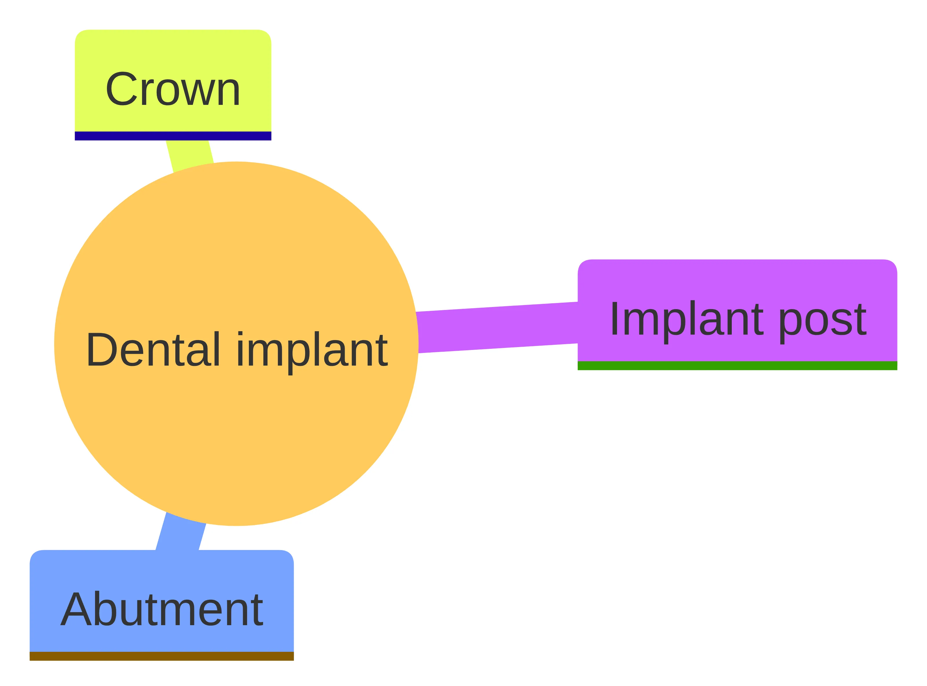
A dental implant has three connected parts that may be described differently in a photograph: the implant post, the abutment, and the crown. The metal post replaces the root portion of a missing tooth. The abutment connects to that post and supports the artificial tooth, or crown.
A close image may show mostly the crown and the gum around it. The post is not necessarily visible in the same view because the implant replaces the root portion beneath the restoration. This distinction matters when you describe what you see. A change in the crown does not automatically show what is happening around the post, and a picture of the gum does not establish whether the underlying implant is stable.
Use neutral descriptions instead of diagnostic labels. For example, note whether the crown appears chipped, shifted, separated from nearby teeth, or different from an earlier image only if that change is genuinely visible. Describe the tissue as red, swollen, or otherwise changed if those features can be seen. Then separate those observations from conclusions such as “the implant failed.”
The useful question is not whether a photograph looks like an example found online. The useful question is which implant part is visible and which findings still require a clinical assessment.
Look for visible changes around the gum
Visible gum changes around a dental implant can provide a reason to seek advice, but they do not identify the cause by themselves. Clinical guidance describes peri-implant health as lacking inflammatory signs such as redness, swelling, and bleeding on probing. A photograph may make redness or swelling noticeable, while bleeding may be reported by you or observed during an examination.
A useful visual review looks for change rather than trying to match a picture to a diagnosis. Compare the concerned area with an earlier image taken from a similar angle and in similar lighting, if you have one. Record what appears different:
- Redness around the gum margin
- Swelling or a change in the gum contour
- Tissue that looks different from the surrounding area
- A visible change in the crown’s position or appearance
It cannot establish whether the tissue change is peri-implant mucositis, peri-implantitis, irritation, or another condition. Peri-implant mucositis affects the mucosa around an implant without involving supporting bone, whereas peri-implantitis also involves supporting bone. Those distinctions require clinical assessment rather than a visual match.
A normal-looking photograph does not prove that the implant is healthy, and a concerning-looking photograph does not prove failure. Treat a visible change as a reason to gather details and seek an evaluation.
Understand what a photograph cannot show beneath the gum
A dental implant photograph cannot show every condition beneath the gum or establish how firmly the implant is attached. The image may capture surface tissue and the crown, but it cannot perform probing, measure mobility, or provide the information that radiographic evaluation may add.
This limitation explains why two images can lead to the wrong conclusion. A picture may show redness without showing the extent of any underlying condition. It may also look unremarkable while you experience movement, pain, bleeding, or swelling. The absence of a dramatic visual finding does not settle a concern that involves stability or symptoms.
Even a series of photographs remains visual information. Comparing images may help show that the appearance changed, but it cannot establish whether supporting bone is involved or whether movement is present. Those questions require the findings gathered during an assessment.
The distinction between visible and nonvisual information is practical:
- Visible information: color, swelling, tissue contour, the portion of the crown shown, and changes between photographs.
- Reported information: pain, bleeding, tenderness, movement, when the concern began, and whether chewing or brushing triggers it.
- Clinical information: probing findings, mobility assessment, and any radiographic findings.
Keep those categories separate when you contact a dentist. Saying “the gum looks red and the area hurts when I chew” gives a fuller account than saying “the implant has failed.” A dentist can then decide which examination steps are appropriate.
Treat mobility, pain, bleeding, and swelling as evaluation prompts
Implant mobility, pain, bleeding, and swelling are reasons to contact a dentist for an assessment, even when a photograph does not look dramatic. These findings can occur with different problems, so they should be recorded as symptoms or observations rather than used as a self-diagnosis.
Mobility deserves particular attention because a cited retrospective review found that 63%–67% of failed implants initially presented with clinical mobility. That percentage describes the failed implants in that retrospective evidence; it does not mean that every mobile-looking crown has the same cause or that a photograph can measure mobility.
Note the details that give the symptom context:
- Whether the implant, crown, or another nearby tooth seems to move
- Whether pain is present and what activity brings it on
- Whether bleeding or swelling is visible or occurs during brushing
- When the symptom began
- Whether the symptom is becoming more noticeable
- Whether chewing, brushing, or temperature triggers it
Do not try to tighten, twist, push, or adjust an implant or loose crown yourself. If a crown feels loose, avoid chewing on that side until a dentist evaluates it.
Why examination and possible imaging matter
A dental examination matters because implant health is assessed through findings that a photograph cannot collect. Peri-implant probing helps assess the tissue around the implant, while a clinician can check mobility and examine the restoration. Radiographic evaluation may add information about structures beneath the gum.
Clinical and radiological findings help distinguish conditions affecting the surrounding mucosa from conditions that also involve supporting bone. The findings can also help a dentist determine what kind of follow-up is appropriate. A picture can support the conversation by showing what first caught your attention, but it cannot supply the examination data itself.
An evaluation can separate several questions that a single image combines: whether the visible tissue appears inflamed, whether the restoration or implant is mobile, and whether supporting bone needs assessment. Those questions call for different clinical findings. The photograph can identify the starting point for the discussion, not answer all of them.
Bring the image to the appointment in its original form if possible. Keep an earlier comparison image, note the date of each photograph, and avoid editing the area in a way that changes its color or shape. Those steps do not make the image diagnostic. They simply help you describe the timeline accurately.
You may not need to know in advance whether imaging will be used. Tell the dentist what you see and feel, then ask what the examination needs to clarify. The clinical assessment, not the visual impression alone, determines what the findings mean.
Put implant failure evidence in clinical context
Implant failure is a clinical conclusion that must be separated from a concerning appearance. Those findings provide context; they do not diagnose your implant.
Peri-implant conditions also have different meanings. Peri-implant mucositis affects the mucosa around an implant without involving supporting bone. Peri-implantitis involves the supporting bone as well. Redness, swelling, bleeding, or pain may prompt assessment, but they do not tell you from a photograph which condition is present.
Published guidance reports higher peri-implantitis risk in people with periodontitis, smokers, implants functioning for more than five years, and people with uncontrolled diabetes or cardiovascular disease. These are factors to mention during a dental history when they apply to you. They are not a substitute for an examination and do not establish that a particular implant has failed.
A large retrospective study cited in the review reported an overall survival rate of 94.4% among 5,532 implants. That result describes one study population and cannot predict the status or future of your implant. Use population evidence to understand why evaluation matters, not to decide what an individual photograph means.
Peri-implant health can also exist around an implant with reduced bone support, according to clinical guidance. That point reinforces the need to keep visual appearance, clinical findings, and interpretation separate.
Record the details a dentist will need
Your symptom timeline and image details can make an implant concern easier to discuss. Before contacting a dentist, write down what changed, when it changed, and what you notice during ordinary activities. The goal is a clear account, not a diagnosis.
Use this short record:
- Location: Identify the implant area and whether the concern is on the upper or lower arch if you know.
- Visible change: Describe redness, swelling, altered tissue contour, or a change in the crown’s appearance.
- Symptoms: Record pain, bleeding, swelling, tenderness, or a sense of movement.
- Timing: Write when you first noticed the concern and whether it is getting worse.
- Triggers: Note whether chewing, brushing, or temperature brings on a symptom.
- Images: Keep the clearest original photograph and any earlier comparison image.
- Relevant history: Mention how long the implant has been functioning and any conditions or habits that your dentist should include in the clinical history.
When you contact a practice, lead with the location, the visible change, the symptoms, and the timeline. For example, describe what you can see and feel rather than opening with a conclusion about failure. You can then ask what information to bring and what the examination needs to clarify.
The symptom details above are useful because dental implant concerns are assessed through more than appearance. A dentist may ask follow-up questions, examine the tissues, check mobility, and decide whether radiographic evaluation is needed.
If you are contacting a practice in South San Francisco, have this record available when you call or complete its contact process. Avoid describing the implant as failed unless a dental professional has made that determination.
Ask focused questions about the implant and crown
Questions about the crown, abutment, implant post, gum tissue, and supporting bone can keep an evaluation focused. A dental implant uses a metal post to replace a missing tooth root, with an abutment supporting the crown. Asking which part may be involved helps you avoid treating the entire restoration as one visible object.
Consider asking:
- Which part of the restoration appears to be involved: the crown, abutment, implant post, or surrounding tissue?
- What clinical findings will you check around the implant?
- Will you assess mobility and probe around the implant?
- Could radiographic evaluation add information that the photograph cannot show?
- What symptoms or changes should prompt me to contact the practice again?
- What should I avoid doing while the concern is being assessed?
- What information from my implant history would help interpret the findings?
These questions do not ask you to choose a treatment from a photograph. They help you understand what the evaluation is intended to clarify. Clinical examination and, when appropriate, radiographic findings are used to distinguish peri-implant conditions and determine the next clinical discussion.
Choose professional evaluation instead of self-diagnosis
Professional evaluation is the appropriate next step when an implant photograph or related symptom concerns you. Contact a dentist, provide the image and symptom record, and describe what changed without assigning a diagnosis. A photograph alone cannot confirm implant failure because assessment may require probing, mobility checks, and radiographic evaluation.
Take these practical steps:
- Save the original photograph and note when it was taken.
- Record pain, bleeding, swelling, movement, timing, and triggers.
- Contact a dental practice and explain the concern clearly.
- Ask what information or examination is needed.
- Avoid tightening, twisting, pushing, or adjusting the implant or crown yourself.
- Avoid chewing on that side if the crown feels loose until it has been evaluated.
Do not wait for a more dramatic image before contacting a dentist if symptoms are worsening or the restoration feels mobile. Prompt dental contact lets the practice determine what evaluation is needed without requiring you to decide what the image means first. If you are in South San Francisco, use the same symptom record when contacting a local dental practice.
What an evaluation can clarify—and what a picture cannot
An implant evaluation can connect your visible observations and symptoms with clinical findings. A dentist may assess the surrounding tissue, probe around the implant, check for mobility, and decide whether radiographic evaluation is appropriate. Those findings can help distinguish inflammation limited to the mucosa from a condition involving supporting bone.
A picture can show the reason you became concerned. It may help communicate redness, swelling, a tissue change, or a difference in the crown’s appearance. It cannot establish the condition of the implant post beneath the gum, measure mobility, or determine whether supporting bone is involved. It also cannot show how symptoms behave during chewing, brushing, or temperature exposure.
Use a simple three-part handoff when you speak with a dentist:
- Observation: Describe what the image shows, such as redness, swelling, a tissue change, or a crown that looks different.
- Experience: Describe pain, bleeding, swelling, tenderness, or movement and identify any triggers.
- Timeline: Explain when you first noticed the change and whether it is worsening.
Keep these conclusions separate:
- A visible change can justify an evaluation. It does not prove failure.
- A symptom can add useful clinical history. It does not identify the cause by itself.
- A stable-looking image can be reassuring as a record. It does not replace an examination.
- Clinical and radiographic findings can add information below the visible surface. They do not turn an online image into a diagnosis.
This approach lets you use photographs responsibly. You can bring useful observations to a dentist while leaving diagnosis, individual suitability, and treatment decisions to the clinical evaluation.
Take the next step with your image and symptom notes
A concerning implant image is a reason to organize your observations, not a reason to diagnose yourself. Keep the photograph, write down your symptoms and timeline, and prepare focused questions about the crown, implant, surrounding tissue, examination, and possible imaging. A qualified dental professional must decide what the findings mean for your individual situation.
If the area is painful, swollen, bleeding, mobile, or changing quickly, communicate those details when arranging care. A dental evaluation is the appropriate way to move from visual concern to an individualized assessment.
What should you do if a dental implant looks concerning in a picture?
What should I do if I notice changes around my dental implant?
If you observe changes such as redness, swelling, or mobility around your dental implant, it’s important to contact your dentist for an evaluation. Document your symptoms, including when they started and any triggers, and provide this information during your appointment.
How can I describe my concerns about a dental implant to my dentist?
When discussing your concerns, describe the visible changes you see, any symptoms like pain or swelling, and the timeline of when you first noticed these changes. This information will help your dentist assess the situation more effectively.
What are the signs that may indicate a dental implant has failed?
Signs that may suggest a dental implant has failed include mobility of the implant, bleeding or swelling around the gum, pain, or changes in the appearance of the crown. However, these signs require professional evaluation for confirmation.
Why is a professional evaluation necessary for dental implant concerns?
A professional evaluation is necessary because a dentist can perform clinical assessments, such as probing and mobility checks, and may use radiographic imaging to gather information that photographs alone cannot provide.
What information should I gather before contacting my dentist about a dental implant?
Before contacting your dentist, gather details about the location of the implant, any visible changes, symptoms you are experiencing, when these changes began, and any relevant medical history that may affect your implant.
Can photographs of dental implants confirm whether they have failed?
No, photographs cannot confirm whether a dental implant has failed. They can provide context for your concerns but cannot replace the clinical findings needed for a proper diagnosis.
What are peri-implant mucositis and peri-implantitis?
Peri-implant mucositis is inflammation of the soft tissue around an implant without affecting the supporting bone, while peri-implantitis involves inflammation that affects both the soft tissue and the supporting bone.
What should I avoid doing if I suspect my dental implant has issues?
Avoid trying to tighten or adjust the implant or crown yourself, and refrain from chewing on that side until a dentist has evaluated the situation.
Need help applying this?
Talk to a billing specialist who knows your payer mix.
Westborough Dental Care welcomes new patients in South San Francisco. Book a visit to meet Dr. Zoulas and the team.
Schedule a meeting