
teeth diseases pictures
Teeth Diseases: What Visible Signs May Suggest
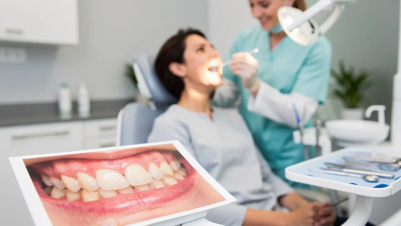
A picture is a clue, not a diagnosis
A picture can help you notice a possible change in a tooth or gum, but it cannot tell you what caused that change. A visual guide is informational; appearance alone does not establish a diagnosis. Pictures may also fail to show symptoms, so a photograph cannot tell you whether you feel pain or how your mouth feels in daily life.
A useful way to look at an image is to separate what you can observe from what you cannot establish. You might notice inflamed, bleeding, or receding gums, or that a tooth looks loose or shifted. A photograph cannot measure gum pockets or show whether bone supporting the teeth has been lost; those findings need a clinical assessment, and an X-ray can show bone loss related to gum disease.
Try describing the concern without naming a disease: note whether the change appears to involve a tooth or gum, what you can see, and whether you also have pain or notice looseness. That description gives you a clear question to bring to a dental visit. It does not turn a picture into a diagnosis.
Treat any match as a reason to pay attention, not proof that the pictured condition is yours. The next sections connect specific visible or felt changes with what they may suggest and what an image leaves unresolved.
Tooth pain can point to different problems
A toothache usually indicates a cavity, but it can also signal gum disease, an abscess, or an impacted tooth. Those possibilities matter because the same report of pain does not identify its cause.
Even if you can point to a particular tooth in an image, its appearance does not settle why it hurts. Avoid assigning a cause from a visual match; the cause needs assessment rather than a guess based on appearance.
A toothache should be evaluated by a dentist right away to determine its cause and help prevent the tooth from dying. When you arrange that evaluation, explain where you feel the pain and what you can see, if anything. Keep the symptom and the image as separate pieces of information: one describes what you feel, and the other shows only what is visible.

Bleeding, inflamed, or receding gums need context
Gum inflammation, bleeding, and recession are changes a dentist may look for during an examination. These are useful words for describing what you notice, but a picture of one change does not establish gum disease or show how extensive it may be.
A gum examination involves more than looking at a photograph. A dentist may use a small probe to measure the spaces around your teeth, called pockets, as well as examine the gums for inflammation, bleeding, or recession. Pocket measurements give information a surface image cannot provide, so do not try to estimate them from a picture.
If you notice bleeding, inflammation, or recession, describe the change plainly at a dental evaluation. You can point out where you see it and mention any other concern you have. Let the examination—not a visual comparison—determine what the finding means for you.
Gum disease can be present without obvious changes
A clear-looking picture cannot rule out gum disease. Gum disease can be present without warning signs, so the absence of an obvious change in an image is not proof that your gums are healthy.
Regular dental checkups and periodontal examinations matter in part because gum disease may have no warning signs. An image gives you a view of visible features; it does not replace those examinations or establish what is happening when there is nothing clear to see.
Loose or shifting teeth warrant professional assessment
A loose or shifting tooth can occur in advanced gum disease. In that setting, destruction of the bone supporting the teeth may cause them to loosen and shift. That association makes the change worth discussing, but it does not prove that gum disease is the cause in your case.
A photograph may show a visible difference in position, but it cannot establish whether a tooth is loose or what is affecting its support. If you notice looseness or shifting, arrange a professional assessment rather than trying to judge the condition from images. An examination may assess loose teeth alongside other clinical findings.
What an examination can reveal beyond a photograph
A periodontal assessment brings together more than visible appearance. Diagnosis follows analysis of medical history, the clinical picture, and symptom examination. Depending on the assessment, those findings may include pocket-depth measurements, an evaluation of loose teeth, radiographic images, blood tests, or a biopsy. The list describes possible parts of an assessment; it is not a set of tests to choose from by looking at a photograph.
Medical history is part of this wider assessment, even though it cannot be seen in a picture. A dentist may ask about health conditions and factors that can contribute to gum disease; tobacco use and diabetes are examples. These details do not prove gum disease or explain a particular visible change by themselves. They provide context to consider alongside the clinical picture and symptoms. An image cannot show these background factors, while knowing about a risk factor cannot substitute for examining the gums, measuring pockets, or checking for bone loss. Each is a different kind of information, and diagnosis draws on the combined assessment rather than a single visible clue.
A dentist may gather information in several ways:
- Examine your gums for inflammation, bleeding, and recession.
- Use a probe to measure the pockets around your teeth.
- Examine your teeth for looseness or shifting.
- Use an X-ray to look for bone loss related to gum disease.
Each step checks a different kind of finding. A picture may help you describe what caught your attention, but it cannot provide pocket measurements or replace an X-ray.
Pocket depth is a measured finding, not a visible feature you can reliably estimate from an image. In a healthy mouth, pockets around the teeth are usually between 1 and 3 millimeters; deeper pockets can be a sign of periodontal disease. The word “can” matters: a measurement is one part of an assessment, not a diagnosis you can make from a photograph.
The 2017 World Workshop criteria describe periodontal health using measured findings: a full-mouth bleeding-on-probing score below 10%, probing depths of 3 millimeters or less, and no clinical attachment loss. Under those same criteria, gingivitis involves bleeding gums at 10% or more of measured sites, with no tooth loss. These are examination measures and definitions; they are not visual thresholds for matching your mouth to a picture.
Read these as combinations of findings. The stated criteria for periodontal health include all three parts: the bleeding score, the pocket-depth limit, and no clinical attachment loss. The gingivitis definition also combines a measured bleeding proportion with the absence of tooth loss. A visible area of bleeding does not show whether the threshold is reached across measured sites or whether the other criteria are met. The percentages and other findings come from clinical measurements, not a visual estimate.
The cited definition of periodontitis also depends on measurements: clinical attachment loss between teeth of 2 millimeters or more at two or more non-adjacent teeth, or attachment loss on the cheek-facing or mouth-facing side of 3 millimeters or more together with pocket depth of 3 millimeters or more at two or more teeth. A picture does not show those measured findings. Keep the figures in their proper role: they describe clinical criteria, not a self-test.
The two routes in this definition are alternatives, not single signs. One requires attachment loss between teeth at multiple non-adjacent teeth; the other requires the stated side-surface attachment loss together with the pocket-depth threshold at multiple teeth. A close-up of one area cannot show whether either pattern of measurements is present across the required teeth. The criteria describe findings a professional assesses, not measurements to attempt from an image.
An X-ray can show whether bone loss has occurred as a result of gum disease. That is different from judging a visible gum change in a photograph. If a picture raises a concern about your gums, an examination can assess findings that the image cannot establish. The examination methods contribute different kinds of information: probing measures pockets, while an X-ray can reveal bone loss. A surface image supplies neither measurement, so a visible change should be considered alongside the other findings rather than treated as a substitute for them.
When a concern should prompt a dental visit
Let the symptom or change guide your next action; do not wait for an image to confirm a disease name. The supported next steps differ by concern:
- Toothache: Contact a dentist for evaluation right away so the cause can be determined.
- Loose or shifting teeth: Arrange a professional assessment; these changes can occur in advanced gum disease.
- Gum changes: Bring inflammation, bleeding, or recession to a dental evaluation. Regular checkups and periodontal examinations also matter because gum disease may have no warning signs.
These points do not assign a severity or diagnosis to an individual sign. They connect a concern with an assessment that can establish what it means.
No visible concern is not a reason to use a picture as a health check. Keep regular dental checkups and periodontal examinations even when you cannot see a change, because gum disease can have no warning signs. A picture can help you explain a concern; it cannot tell you that no concern exists.
Before your visit, put your concern into simple terms: what you see, whether you feel pain, and whether you notice a tooth becoming loose or shifting. You do not need to label the condition. A clear description helps keep the conversation focused on the signs you noticed and the examination needed to assess them.
Use pictures to guide your next conversation with a dentist
A picture is a starting point for describing what you notice, not a way to diagnose a condition or judge its severity. Only a dentist, physician, or other qualified health care professional can make a diagnosis. Bring your visible concern and symptoms into the conversation, and leave the interpretation to an individual assessment.
Questions about interpreting teeth and gum changes
What does stage 1 periodontitis look like in pictures?
A picture cannot establish whether gum disease is at an early stage. Assessment relies on clinical findings such as gum-pocket measurements and other examination information; a dentist can interpret those findings in context.
Can a photo show whether gum disease has caused bone loss?
A surface photo cannot establish bone loss. A dentist may use an X-ray to look for bone loss related to gum disease, alongside an examination and other relevant findings.
What should I do if a tooth looks different but does not hurt?
A change in appearance alone does not identify its cause. Note what you see and mention it at a dental evaluation; a photograph cannot rule out a problem or confirm a diagnosis.
Need help applying this?
Talk to a billing specialist who knows your payer mix.
Westborough Dental Care welcomes new patients in South San Francisco. Book a visit to meet Dr. Zoulas and the team.
Schedule a meeting